You have successfully logged out.
Not registered yet?
Help prevent and treat diabetic foot ulcers
Risk of diabetic foot syndrome
Diabetic foot syndrome (DFS) is a common complication of diabetes mellitus that affects every fourth patient with diabetes at some point in their life.[1] The risk of developing DFS increases with time.
On this page
Medical Professional
This information is meant for medical professionals only. Please confirm that you are a medical professional before accessing the information.
Confirm Yes, I am a health care professional. Cancel No, I am not a health care professional.Possible consequences: Diabetic foot ulcer (DFU) or Charcot foot
One consequence of DFS is an ultimate skin breakdown or foot ulceration, which includes all changes in the foot as a result of diabetic polyneuropathy as well as diabetic micro- and macroangiopathic changes.
Patients may also develop Charcot foot, which is another manifestation of DFS. Charcot foot is a deformation characterized by a deterioration in the bone metabolism due to diabetes, which results in fractures of the bones, joint dislocations, fusion of bone fragments, and other complex deformations of the foot. Another name for Charcot foot is diabetic osteoarthropathy.
Diabetic Charcot foot may develop without a foot ulcer, but it is often associated with diabetic foot ulcers. The deformities lead to pressure ulcers because of standing or walking, or simply when patients wear shoes that are not designed for their specific foot anatomy.[1,2]
Diabetes can affect the peripheral nervous system in many ways. Patients often experience numbness, tingling, pain, and/or weakness that start in the feet and increase the risk of foot ulcers. This is caused by tight shoes, abrasive socks, or unnoticed microtrauma to the skin. Moreover, the skin’s resistance to trauma may be impaired because the function of skin appendages, including hair follicles and sebaceous glands, is altered in diabetic neuropathy.[3,4]
Another risk factor that contributes to DFS is diabetic angiopathy. Larger arteries stiffen due to fibrosis, media enlargement, calcification and multiple other processes (macroangiopathy). But also small vessels, especially the inner layer of capillaries are affected by diabetes comprising their function (microangiopathy). Both can critically contribute to DFS.[1,5,6,7]
Skin integrity of the foot
Help to prevent the formation of diabetes foot ulcers

Description
No open lesion; patient may have severely deformed foot.
Goal
Maintain skin integrity

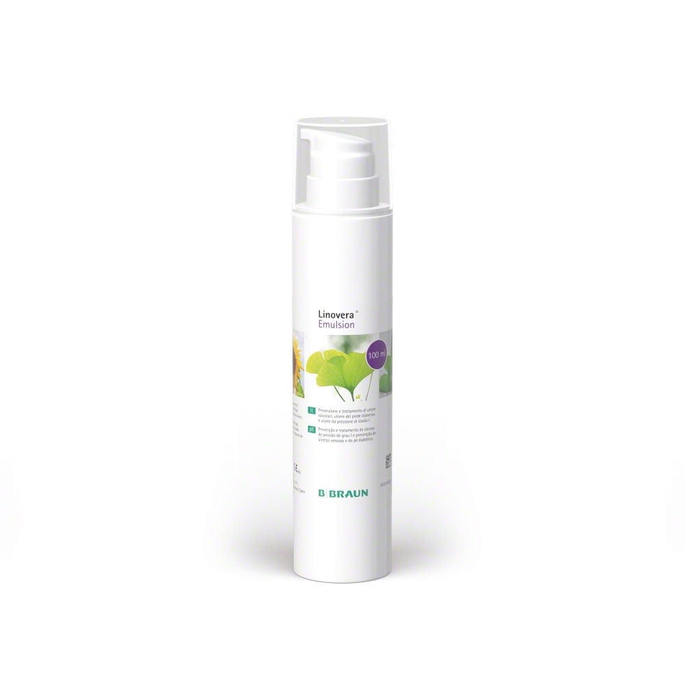
The formulation of Linovera® and Linovera® Emulsion is based on hyperoxygenated fatty acids, important linoleic acids which can contribute to a healthy skin structure. Linovera® Emulsion is indicated for prevention and treatment of lower limb ulcers, diabetic foot ulcers and stage 1 pressure ulcers.

Protect from friction
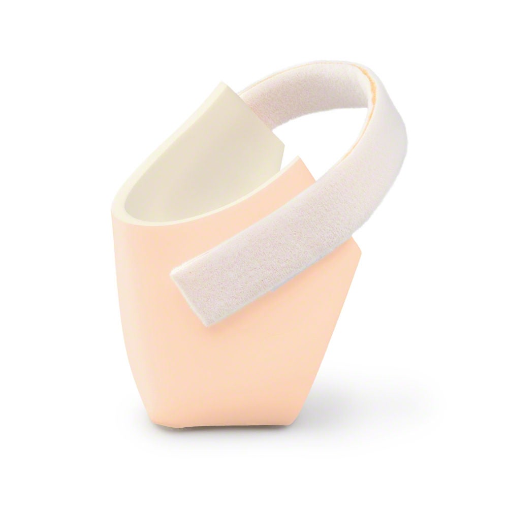
Askina® Heel is a non adhesive hydrocellular heel dressing that protects the heel area from shear stresses and reduces pressure from external forces.
Management of diabetes foot ulcers
A top priority in treating the diabetic foot syndrome is to avoid amputation.[8] Because currently, the majority of all foot and lower leg amputations are performed on patients with diabetes mellitus. It is estimated that amputations occur 40 times more frequent in patients affected by diabetes compared to non-diabetic patients.[9]
Treatment guide
Depending on the extent of the tissue damage, diabetic foot ulcers can be categorized into five grades, with every grade requiring its specific treatment.[10,11,12]

Description
Superficial ulcer involving the full skin thickness but not underlying tissues
Goal
Keep the wound bed clean for granulation tissue to form
Manage biofilm
Prontosan® Wound Irrigation Solution is indicated for cleansing irrigation and moistening of acute and chronic wounds.
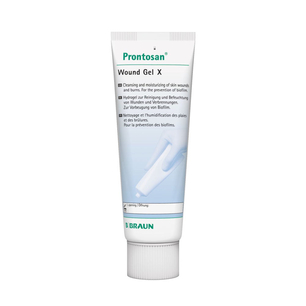
The use of Prontosan® Wound Gel X can provide long-lasting cleansing and decontamination of the wound bed between dressing changes.
Prontosan® Debridement Pad has been designed to support the wound bed preparation when used in conjunction with Prontosan® Wound Irrigation Solution.

Manage wound exudate
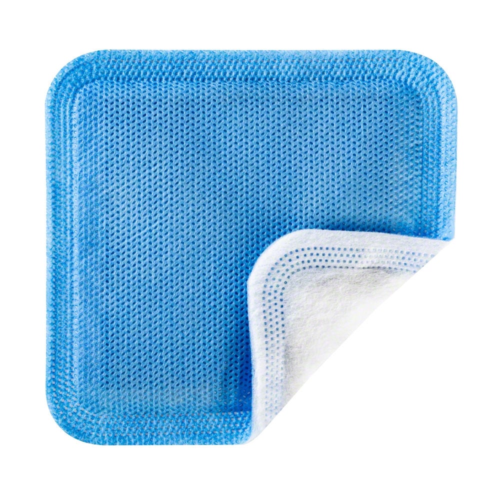
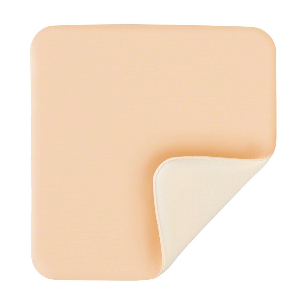
Askina® DresSil Border is a self adherent foam dressing with soft silicone adhesive on one side and a vapor permeable waterproof film on the other. With its circular shaped island of foam and the additional adhesive border it is specially designed for small, deep wounds, e.g. diabetic foot ulcers.


Description
Deep ulcer that penetrates down to the ligaments and muscles but not to the bones.
Goal
- Remove slough/callus
- Prevent/remove biofilm
- Reduce bacterial load
- Manage exudate
Manage biofilm
Prontosan® Wound Irrigation Solution is indicated for cleansing irrigation and moistening of acute and chronic wounds.
The use of Prontosan® Wound Gel X can provide long-lasting cleansing and decontamination of the wound bed between dressing changes.
Prontosan® Debridement Pad has been designed to support the wound bed preparation when used in conjunction with Prontosan® Wound Irrigation Solution.

Manage wound exudate
Askina® DresSil and Askina® DresSil Border help to maintain a moist wound environment conducive to natural healing conditions with a perfored silicone wound contact layer, a highly absorbent polyurethane foam and a vapor permeable waterproof outer film.


Description
Deep ulcer with cellulitis or abscess formation, often with osteomyelitis
Goal
- Remove slough/callus
- Prevent/remove biofilm
- Reduce bacterial load
- Manage exudate/odor
Manage biofilm
Prontosan® Wound Irrigation Solution is indicated for cleansing irrigation and moistening of acute and chronic wounds.
The use of Prontosan® Wound Gel X can provide long-lasting cleansing and decontamination of the wound bed between dressing changes.
Prontosan® Debridement Pad has been designed to support the wound bed preparation when used in conjunction with Prontosan® Wound Irrigation Solution.

Manage wound exudate
Askina® Foam absorbs wound exudate through its polyurethane foam providing a moist wound healing environment. It has a vapor permeable, water and bacteria resistant polyurethane film outer layer.


Manage wound odor
Askina® Carbosorb is an activated charcoal super absorbent dressing, suitable for moderate to heavily exuding wounds.

Description
Localized gangrene
Goal
- Remove slough/callus
- Prevent/remove biofilm
- Reduce bacterial load
- Manage exudate/odor
Manage biofilm
Prontosan® Wound Irrigation Solution is indicated for cleansing irrigation and moistening of acute and chronic wounds.
The use of Prontosan® Wound Gel X can provide long-lasting cleansing and decontamination of the wound bed between dressing changes.
Prontosan® Debridement Pad has been designed to support the wound bed preparation when used in conjunction with Prontosan® Wound Irrigation Solution.

Manage wound exudate
Askina® Foam absorbs wound exudate through its polyurethane foam providing a moist wound healing environment. It has a vapor permeable, water and bacteria resistant polyurethane film outer layer.


Manage wound odor
Askina® Carbosorb is an activated charcoal super absorbent dressing, suitable for moderate to heavily exuding wounds.
Our product portfolio
Skin prevention and protection
Wound bed preparation and biofilm management
Wound infection and odor management
Exudate management
[1] Rümenapf G, Morbach S, Rother U, Uhl C, Görtz H, Böckler D, Behrendt CA, Hochlenert D, Engels G, Sigl M; Kommission PAVK und Diabetisches Fußsyndrom der DGG e. V.. Diabetisches Fußsyndrom – Teil 1 : Definition, Pathophysiologie, Diagnostik und Klassifikation [Diabetic foot syndrome-Part 1 : Definition, pathophysiology, diagnostics and classification]. Chirurg. 2021 Jan;92(1):81-94. German. doi: 10.1007/s00104-020-01301-9. PMID: 33170315; PMCID: PMC7819949.
[2] Sommer TC, Lee TH. Charcot foot: the diagnostic dilemma. Am Fam Physician. 2001 Nov 1;64(9):1591-8. Erratum in: Am Fam Physician 2002 Jun 15;65(12):2436-8. PMID: 11730314.
[3] Callaghan BC, Cheng HT, Stables CL, Smith AL, Feldman EL. Diabetic neuropathy: clinical manifestations and current treatments. Lancet Neurol. 2012 Jun;11(6):521-34. doi: 10.1016/S1474-4422(12)70065-0. Epub 2012 May 16. PMID: 22608666; PMCID: PMC4254767.
[4] Takeo M, Lee W, Ito M. Wound healing and skin regeneration. Cold Spring Harb Perspect Med. 2015 Jan 5;5(1):a023267. doi: 10.1101/cshperspect.a023267. PMID: 25561722; PMCID: PMC4292081.
[5] Lauterbach S, Kostev K, Kohlmann T. Prevalence of diabetic foot syndrome and its risk factors in the UK. J Wound Care. 2010 Aug;19(8):333-7. doi: 10.12968/jowc.2010.19.8.77711. PMID: 20852505
[6] Sämann A, Tajiyeva O, Müller N, Tschauner T, Hoyer H, Wolf G, Müller UA. Prevalence of the diabetic foot syndrome at the primary care level in Germany: a cross-sectional study. Diabet Med. 2008 May;25(5):557-63. doi: 10.1111/j.1464-5491.2008.02435.x. Epub 2008 Mar 14. PMID: 18346154.
[7] Strain WD, Paldánius PM. Diabetes, cardiovascular disease and the microcirculation. Cardiovasc Diabetol. 2018 Apr 18;17(1):57. doi: 10.1186/s12933-018-0703-2. Erratum in: Cardiovasc Diabetol. 2021 Jun 11;20(1):120. PMID: 29669543; PMCID: PMC5905152.
[8] Olm M, Kühnl A, Knipfer E, Salvermoser M, Eckstein HH, Zimmermann A. Operative Versorgung von Diabetikern mit vaskulären Komplikationen : Sekundärdatenanalyse der DRG-Statistik von 2005 bis 2014 in Deutschland [Operative treatment of diabetics with vascular complications : Secondary data analysis of diagnosis-related groups statistics from 2005 to 2014 in Germany]. Chirurg. 2018 Jul;89(7):545-551. German. doi: 10.1007/s00104-018-0628-z. PMID: 29589075
[9] Narres M, Kvitkina T, Claessen H, Droste S, Schuster B, Morbach S, Rümenapf G, Van Acker K, Icks A. Incidence of lower extremity amputations in the diabetic compared with the non-diabetic population: A systematic review. PLoS One. 2017 Aug 28;12(8):e0182081. doi: 10.1371/journal.pone.0182081. PMID: 28846690; PMCID: PMC5573217.
[10] Jeon BJ, Choi HJ, Kang JS, Tak MS, Park ES. Comparison of five systems of classification of diabetic foot ulcers and predictive factors for amputation. Int Wound J. 2017 Jun;14(3):537-545. doi: 10.1111/iwj.12642. Epub 2016 Oct 10. PMID: 27723246; PMCID: PMC7949506.
[11] Santema TB, Lenselink EA, Balm R, Ubbink DT. Comparing the Meggitt-Wagner and the University of Texas wound classification systems for diabetic foot ulcers: inter-observer analyses. Int Wound J. 2016 Dec;13(6):1137-1141. doi: 10.1111/iwj.12429. Epub 2015 Feb 26. PMID: 25720543; PMCID: PMC7949589.
[12] Wagner FW. The dysvascular foot: a system for diagnosis and treatment. Foot Ankle 1981;2
Stay connected with My B. Braun
With your personalized account, your online experience will be easier, more comfortable and safe.
