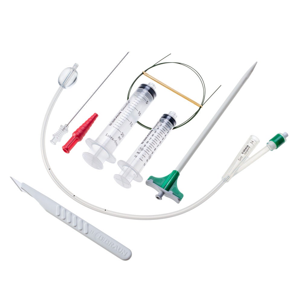
Cystofix® SG set
Cystofix® SG set: Puncture set for suprapubic catheterization
The Cystofix® SG Set product comprises a sterile, single use, Seldinger Technique set for suprapubic bladder drainage.
Advantages
Sterility
- Cystofix® is a range of sterile, state-of-the-art catheters and catheter placement / exchange accessories for suprapubic catheterisation.1, 2, 3
Patient Comfort
- Depending on circumstances, suprapubic catheterization can be the preferred method of indwelling urinary catheterization, particularly in terms of incidence of asymptomatic bacteriuria and patient comfort.1
- The silicone catheters from the Cystofix® SG range have incorporated balloons.4
- Short tip silicone balloon catheters such as those of the Cystofix® provide increased patient comfort compared to balloon catheters with a long extremity.4
- Cystofix® SG uses a placement method (Seldinger technique) where the initial puncture of the abdominal wall is made with an 18G needle whose diameter is much less than that of a splitable cannula.5
Usage Time
- The biological tolerability of the silicone material used for the Cystofix® balloon catheters allow for durations of use of up to 30 days and for replacement as many times as clinically needed.4
Balloon Catheter
- Made of silicone to allow a usage up to 30 days
- Printings from the catheter enable the user to determine the catheter's position in the bladder or in the cannula, during puncturing
- Available with 40 cm length (straight tip)
- Open tip to enable the use of a guidewire during the replacement
- For easier removal with reduced risk of cuffing
- For easier insertion at catheter replacement (no ridge)
- Y-shaped connector, with anti-reflux valve and Luer-Lock fitting
- Can be replaced repeatedly as appropriate over long periods of time
Guidewire
- Made of stainless-steel with PTFE (Polytetrafluoroethylene)
- Used for Seldinger Technique method, allows a path for the catheter insertion after dilator removal
- Stiff shaft
- Straight and flexible tip
Dilator
- For Cystofix® SG, enables the user to dilate the suprapubic path after guide wire positioning
- Smooth color-coded shape dilator available from CH10 to CH16
Introducer
- For Cystofix SG, allows a path for the catheter insertion after dilator remova
- Introducer wings enable the user to split the introducer by halves for removal
Stopper
- To stop the urine flow on demand
Puncture Needle
- Enables the user to puncture the abdominal wall till the bladder
- Made of stainless-steel with 18G and 12cm length
- Used for Seldinger Technique method, allows a path for the guidewire insertion after the local injected anesthetic
Scalpel
- Enables the user to cut the skin before the abdominal wall puncture
Syringes 10 and 20 ml
- 20ml syringe used to inject anesthesia through abdominal wall till the bladder
- 10ml syringe used to inflate the balloon of the catheter
Therapy application
- Suprapubic catheterization is a bladder management method where a catheter is inserted through the abdominal wall into the bladder and maintained in place for continuous urinary drainage.
- The Cystofix® SG product range is intended to be used for the drainage of the bladder through the abdominal wall, on the median line of the body above the symphysis (puncture set).
- The catheter is introduced in the bladder to allow the drainage after surgery, in case of bladder dysfunction or urinary retention.
- E. A. Kidd, F. Stewart, N. C. Kassis, E. Hom, M. I. Omar; “Urethral (indwelling or intermittent) or suprapubic routes for short-term catheterisation in hospitalized adults” Cochrane Database Syst Rev. 2015 December 10.
- A. Kulbay, E. Joelsson-Alm, A. Tammelin; “The impact of guidelines on sterility precautions during indwelling urethral catheterization at two acute-care hospitals in Sweden - a descriptive survey” European Association of Urology Nurses (EAUN).
- British Association of Urological Surgeons’ guidelines (BAUS) - Hall et al. 2020, Harrison et al. 2011.
- Data on file.
- “Seldinger SI. Catheter replacement of the needle in percutaneous arteriography; a new technique.” Acta radiologica. 1953; 39 (5): 368–76.


